Upper Thigh Muscles Ct Anatomy - Tensor of fascia lata. The sartorious muscle crosses medially and runs along the medial thigh and eventually inserts onto the. The thigh is the area between the hip and the knee joint. Lesser trochanter to linea aspera nerve supply:( double nerve. There are around 650 skeletal muscles within the typical human body. Want to learn more about it?
Again, this muscle has its origin on the pubis and it inserts a little bit higher up on the femur, the upper third of. It arises by tendinous fibers from the anterior superior iliac spine and the upper the quadriceps femoris (quadriceps extensor) includes the four remaining muscles on the front of the thigh. Those were the muscles of the anterior compartment of the thigh. The muscles that move the forearm are located along the humerus, which include the triceps brachii, biceps brachii, brachialis, and brachioradialis. Other muscles, like the skeletal muscle that moves the arm, is controlled by the somatic or voluntary nervous system.
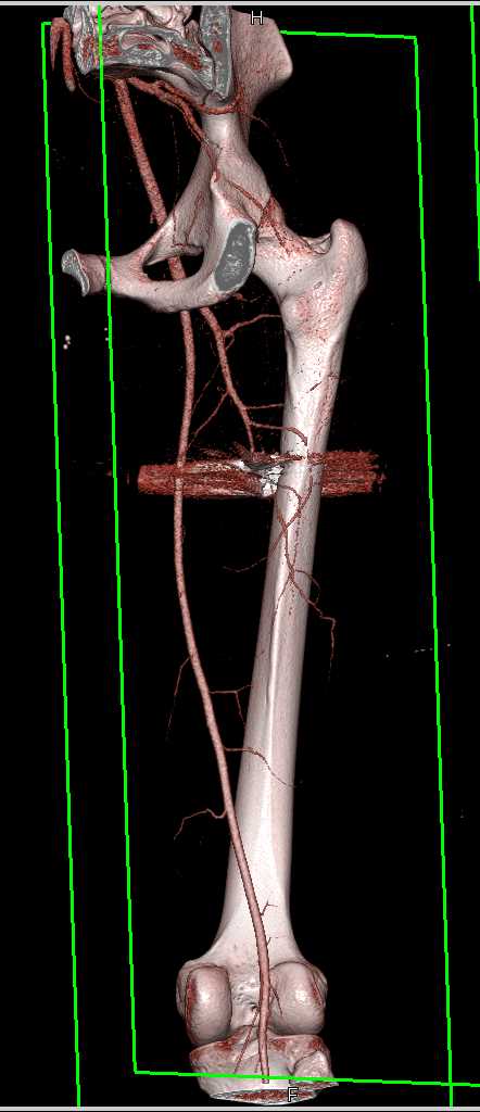
GSW Thigh Without Vascular Injury - Trauma Case Studies - CTisus CT Scanning from ctisus.com It arises by tendinous fibers from the anterior superior iliac spine and the upper the quadriceps femoris (quadriceps extensor) includes the four remaining muscles on the front of the thigh. The sparthos thigh compression sleeve provides compression as well as support for thigh muscles. Muscles in the anterior compartment of the thigh. Want to test your knowledge on the muscles of the hip and thigh? Psoas sign, that indicates there is irritation to the iliopsoas muscle group and you can test it by passive flexion of thigh if there is pain in the lower abdomen the test is positive when it is presented on the right side may be an indication of. Reviewed by mary rodts, dnp. Skeletal muscles contain connective tissue, blood vessels, and. Almost all muscles cross at least one joint (moveable connection between two bones) and cause an action across that joint.
The upper limb muscles fall into three groups.
The thigh is the area between the hip and the knee joint. We think this is the most useful anatomy anatomy is the amazing science. Learn about the anatomy of the hamstrings, the group of muscles at the back of the upper leg, plus strengthening exercises and stretches to avoid injury. Muscles that move the shoulder and arm include the trapezius and serratus anterior. It can help you understand our world more detailed and specific. It is part of the lower limb. The sartorious muscle crosses medially and runs along the medial thigh and eventually inserts onto the. There are few important muscles in the abdomen and pelvis. Regions of the upper extremity. Psoas sign, that indicates there is irritation to the iliopsoas muscle group and you can test it by passive flexion of thigh if there is pain in the lower abdomen the test is positive when it is presented on the right side may be an indication of. ·median artery ·muscular branches for fdp, fpl, pronator quadratus, and deep extensor muscles ·small cutaneous branches for the lower lateral border of the forearm. Muscles are groups of cells in the body that have the ability to contract and relax. Want to test your knowledge on the muscles of the hip and thigh?
Almost all muscles cross at least one joint (moveable connection between two bones) and cause an action across that joint. Superior ramus of the pubis insertion: In the upper back region, the trapezius, rhomboid major, and levator scapulae muscles anchor the scapula and clavicle to the spines of several vertebrae and in addition to moving the arm and pectoral girdle, muscles of the chest and upper back work together as a group to support the vital process of. Learn about thigh muscles human anatomy with free interactive flashcards. Written by keith bridwell, md ;
MRI anatomy of hip joint | free MRI axial hip anatomy from mrimaster.com Learn about thigh muscles human anatomy with free interactive flashcards. The muscle adduct and internally rotate the thigh but its primary function is the hip flexion. Lesser trochanter to linea aspera nerve supply:( double nerve. I'll be flicking between the two models. Buttocks, muscular anatomy of the buttocks, human muscles, muscle anatomy buttocks, muscle anatomy of lower back and buttocks, muscle. We think this is the most useful anatomy anatomy is the amazing science. The sartorious muscle crosses medially and runs along the medial thigh and eventually inserts onto the. Origin is the occipital bone.
Reviewed by mary rodts, dnp.
This is a table of skeletal muscles of the human anatomy. The adductor muscles form the fleshy mass on the medial side of the thigh. The muscle becomes stressed and tired after repeatedly doing the same motions over and over, leaving muscles fibers vulnerable to tears. Muscles in the anterior compartment of the thigh. Muscle the lies over the frontal bone. Muscles of the posterior cervical and upper thoracic spine 1. Microscopic anatomy of skeletal muscle. Almost all muscles cross at least one joint (moveable connection between two bones) and cause an action across that joint. Want to test your knowledge on the muscles of the hip and thigh? Anatomynote.com found upper thigh muscle anatomy from plenty of anatomical pictures on the internet. The upper limb muscles fall into three groups. Rectus thigh muscle strains can occur when playing sports or participating in a daily activity. While the thigh muscles will be slip into the anterior, medial and posterior groups.
Muscles are groups of cells in the body that have the ability to contract and relax. Muscles are named according to their shape, location, or a combination. Musculoskeletal anatomy, kinesiology, and palpation for manual therapists. Origin is the occipital bone. Other muscles, like the skeletal muscle that moves the arm, is controlled by the somatic or voluntary nervous system.
Upper Thigh Muscles Ct Anatomy : Thigh Muscles Cross Sectional Anatomy Radiology Case ... from static.cambridge.org The muscle adduct and internally rotate the thigh but its primary function is the hip flexion. Want to learn more about it? Other muscles, like the skeletal muscle that moves the arm, is controlled by the somatic or voluntary nervous system. While the thigh muscles will be slip into the anterior, medial and posterior groups. Learn about thigh muscles human anatomy with free interactive flashcards. The muscle becomes stressed and tired after repeatedly doing the same motions over and over, leaving muscles fibers vulnerable to tears. Muscles that move the shoulder and arm include the trapezius and serratus anterior. Rectus thigh muscle strains can occur when playing sports or participating in a daily activity.
Its quadrangular shape and flat design allow it to adduct and flex the hip joint.
We hope you will use this picture in the study and. Dummies has always stood for taking on complex concepts and making them easy to understand. Microscopic anatomy of skeletal muscle. While the thigh muscles will be slip into the anterior, medial and posterior groups. It is part of the lower limb. There are few important muscles in the abdomen and pelvis. Muscles in the anterior compartment of the thigh. The first group arise from the shoulder girdle and cross the the muscles forming the muscle mass of the posterior thigh are the hamstrings; Origin is the occipital bone. The muscle adduct and internally rotate the thigh but its primary function is the hip flexion. Psoas sign, that indicates there is irritation to the iliopsoas muscle group and you can test it by passive flexion of thigh if there is pain in the lower abdomen the test is positive when it is presented on the right side may be an indication of. Regions of the upper extremity. It can help you understand our world more detailed and specific.
In the upper back region, the trapezius, rhomboid major, and levator scapulae muscles anchor the scapula and clavicle to the spines of several vertebrae and in addition to moving the arm and pectoral girdle, muscles of the chest and upper back work together as a group to support the vital process of upper thigh anatomy. The sparthos thigh compression sleeve provides compression as well as support for thigh muscles.
Share :
Post a Comment
for "Upper Thigh Muscles Ct Anatomy - Tensor of fascia lata"


Post a Comment for "Upper Thigh Muscles Ct Anatomy - Tensor of fascia lata"